|
|
|
|
|
|||||||||||
|
Electronic Flora of South Australia Species Fact Sheet
Phylum Rhodophyta – Family Delesseriaceae
Thallus (Fig. 51A) foliose, usually simple, rose-red to dark purple, 10–35 cm high and 5–20 cm broad, margins smooth to densely ruffled, with a slender, simple or branched, stipe 1–2 (–4) cm long and 0.5–1 mm broad; macroscopic veins very faint, simple or subdichotomous, present for 5–10 cm above the stipe, 0.5–2 mm broad. Holdfast discoid, lobed; epizoic on gastropods or bivalves. Structure. Growth of young blades by apical cells of marginal dentations, later by vague marginal meristems, with scattered intercalary divisions. Blades monostromatic except for veins (Fig. 51B) and reproductive areas, cells more-or-less isodiametric, 20–60 µm across in surface view. Veins developed by periclinal divisions of linear series of cells, followed by anticlinal and further periclinal divisions, mature veins 20–25 cells thick. Cells multinucleate, nuclei in a medial horizontal layer; rhodoplasts one to a few per cell, lobed, often ribbon like.
Reproduction: Gametophytes dioecious. Procarps scattered, with the primary cells cutting off pericentral cells (usually on both sides of the blade) which act as supporting cells, bearing 2 one-celled sterile groups and a 4-celled carpogonial branch, but without a cover cell. Carposporophyte(Fig. 51C) developing a massive, branched, fusion cell and much branched gonimoblast bearing pyriform terminal carposporangia 40–65 µm in diameter, with lower cells also maturing as carposporangia. Cystocarps protuberant, 0.5–0.8 mm across; pericarp ostiolate (often slightly eccentric), 40–75 µm and 2–4 cells thick. Spermatangial sori (Fig. 51D) scattered, usually minute, irregular in shape, sometimes coalescing, with the primary cells dividing once periclinally, then anticlinally to form a group of initials each of which cuts off 2–4 spermatangia.
Tetrasporangial sori scattered, ovate, 200–550 µm across and 180–220 1 µm thick, with tetrasporangia in 2 layers (Fig. 51E), cut off from inner cortical cells, subspherical (Fig. 51F), (45–) 90–120 µm in diameter.
Type from Ninepin Point, SE Tasmania, 9–12 m deep (Kraft & Sanderson, 22.xii.1992; holotype MELU, K9167 (cystocarpic), syntype K9135 (tetrasporangial). Isotypes in MELU and in AD, A68023, A68024.
Selected specimens: Ninepin Point, Tas., 9–15 m deep (Kraft & Sanderson, 4.i.1993; MELU, 42219, K9197). Arch Rock, Tas., 6–15 m deep (Kraft & Scott, 16.xii.1993; MELU, K9855). 2 km N of Satellite I., D'Entrecasteaux Ch., Tas., on gastropod Maoricolpus, 15 m and 12 m deep (Shepherd, 17.ii.1972; AD, A41663 and A41668 resp.)
Distribution: Only known from the type locality and nearby Arch Rock and from 2 km N of Satellite I., D'Entrecasteaux Ch., Tasmania.
Taxonomic notes: S. tasmanica is a particularly fine species known mainly from an extensive suite of specimens collected by Kraft & Sanderson from Ninepin Point.
References:
LIN, S.-M. & KRAFT, G.T. (1999). Schizoseris tasmanica sp. nov. (Delesseriaceae, Ceramiales), a first record of the genus for the Australian marine flora. Phycologia 38, 128–137.
The Marine Benthic Flora of Southern Australia Part IIID complete list of references.
Publication:
Womersley, H.B.S. (24 February, 2003)
The Marine Benthic Flora of Southern Australia
Rhodophyta. Part IIID. Ceramiales – Delesseriaceae, Sarcomeniaceae, Rhodomelaceae
Reproduced with permission from The Marine Benthic Flora of Southern Australia Part IIID 2003, by H.B.S. Womersley. Australian Biological Resources Study, Canberra. Copyright Commonwealth of Australia.
Illustration in Womersley Part IIIA, 2003: FIG. 51.
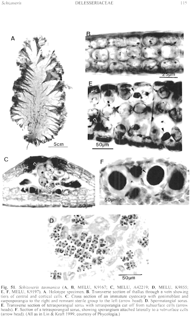
Figure 51 enlarge
Fig. 51. Schizoseris tasmanica (A, B, MELU, K9167; C, MELU, A42219; D, MELU, K9855; E, F, MELU, K9197). A. Holotype specimen. B. Transverse section of thallus through a vein showing tiers of central and cortical cells. C. Cross section of an immature cystocarp with gonimoblast and carposporangia to the right and remnant sterile group to the left (arrow head). D. Spermatangial sorus. E. Transverse section of tetrasporangial sorus with tetrasporangia cut off from subsurface cells (arrow heads). F. Section of a tetrasporangial sorus, showing sporangium attached laterally to a subsurface cells (arrow head). (All as in Lin & Kraft 1999, courtesy of Phycologia.)

|
Email Contact: State Herbarium of South Australia |

|