|
|
|
|
|
|||||||||||
|
Electronic Flora of South Australia Species Fact Sheet
Phylum Rhodophyta – Family Rhodomelaceae – Tribe Lophothalieae
Selected citations: J. Agardh 1890: 61. De Toni 1903: 1018. Falkenberg 1901: 534, pl. 13 figs 24–28. Guiler 1952: 104. Kylin 1956: 511. Lucas 1909: 43; 1929a: 22; 1929b: 51. Lucas & Perrin 1947: 285. May 1965: 378. Parsons 1975: 653, figs 28, 29, 41 B. Reinbold 1897: 57. Schmitz 1893: 219. Wilson 1892: 166. Womersley 1966: 153.
Synonym
Dasya verticillata Harvey 1844b: 434; 1846: 422; 1847: 64, pl. 24; 1859b: 304; 1863, synop.: xxiv. J. Agardh 1863: 1234; 1890: 61. Sonder 1853: 702; 1880: 36.
Thallus (Fig. 119A) medium to dark red-brown, fading to grey-red, 10–30 (–50) cm high, much branched with robust, cartilaginous, usually branched main axes (1–) 2–3 mm in diameter, bearing delicate, slender, irregular, terete laterals for 3–5 orders, basally 0.5–1 mm in diameter, tapering to 50–100 µm in diameter below apices; cortication commencing near apices but slight on laterals, becoming thick and dense on axes. Laterals clothed throughout (Fig. 119B) with slender, simple, rhodoplastic trichoblasts (one per axial cell) and monosiphonous adventitious filaments (Fig. 120A) arising from the pericentral cells more or less in a whorl. Holdfast divided, fibrous, 3–10 (–15) mm across; epilithic or on jetty piles. Structure monopodial, with axial cells producing 5 pericentral cells in alternating sequence, sometimes with 1 or 2 further intercalated cells, pericentral cells usually clear in transverse section (Fig. 119C). Trichoblasts 30–50 (–75) µm in diameter with basal cells L/D 1–2, increasing above to L/D 3–8 (–20); adventitious filaments similar but slightly more slender. Lateral branches arising on lower cells of trichoblasts. Cells uninucleate; rhodoplasts discoid.
Reproduction: Gametophytes dioecious. Procarps (Fig. 120B) arising 4–7 cells from base of trichoblasts, with 5 pericentral cells, the last-formed cutting off a sterile group initial (becoming 3-celled) then the initial of the 4-celled carpogonial branch. Carposporophyte (Fig. 120C) with a broad to erect fusion cell with short, branched, gonimoblast filaments bearing clavate terminal carposporangia 30–45 µm in diameter, replaced from lower cells. Cystocarps (Figs 119D, 120D) urceolate with a short stalk and slight neck, 1–1.5 mm in diameter; pericarp ostiolate, 2 cells thick. Spermatangial organs (Fig. 119E) covering the basal part of the trichoblasts with a sterile upper filament, elongate, 180–250 µm long and 30–50 µm in diameter.
Tetrasporangial stichidia (Fig. 119F) transformed from vegetative branches, 0.5–1.5 mm long and 100–180 µm in diameter, bearing trichoblasts, with decussate pairs (Fig. 120E) of tetrasporangia 50–80 µm in diameter, each with 2 pre-sporangial and 1 post-sporangial cover cells.
Type from Georgetown, Tas.; holotype Gunn 1306 in BM.
Selected specimens: Outside Tapley Shoal (Edithburg), S. Aust., 15 m deep (Shepherd, 2.ii.1969; AD, A33500). Aldinga, S. Aust., drift (Womersley, 17.ix.1966; AD, A30712-data in Parsons 1975, p. 653 incorrect). Granite I. causeway, Victor Harbor, S. Aust., 7–10 m deep (Edyvane, 8.viii.1982; AD, A53198). Vivonne Bay, Kangaroo I., S. Aust., 2–4 m deep on jetty pylons (Kraft & Min-Thein, 4.xii.1971; AD, A41396). Penneshaw, Kangaroo I., S. Aust., on rope on jetty, 7 m deep (Lavers, 16.ix.1996; AD, A66795). Stinky Bay Point, Nora Creina, S. Aust., 3–5 m deep (Millar, 28.x.1996; AD, A67205). St Leonards, Port Phillip, Vic., 1–3 m deep (Womersley, 9.viii.1959; AD, A23095). Port Phillip, Vic., 17 m deep, N bay (Macpherson, 18.x.1959; AD, A24659). San Remo, Vic. drift on outer beach (Sinkora A1964, 27.xi.1974; AD, A55598). Low Head, Tas. (Perrin, 11.xi.1950; AD, A16496). Bridport, Tas., drift (Womersley & Parsons, 6.xi.1982; AD, A54547). Mussleroe Bay, Tas. (Perrin, Mar. 1950; AD, A49964).
Distribution: Tapley Shoal, S. Aust., to San Remo, Vic., and N Tasmania.
Taxonomic notes: L. verticillata was described in detail by Parsons (1975, p. 653). It differs from L. hormoclados in developing whorled trichoblasts and copious adventitious filaments and in their distinctly smaller cells.
References:
AGARDH, J.G. (1863). Species Genera et Ordines Algarum. Vol. 2, Part 3, pp. 787–1291. (Gleerup: Lund.)
AGARDH, J.G. (1890). Till algernes systematik. Acta Univ. lund. 26(3), 1–125, Plates 1–3.
DE TONI, G.B. (1903). Sylloge Algarum omnium hucusque Cognitarum. Vol. 4. Florideae. Sect. 3. pp. 775–1521 + 1523–1525. (Padua.)
FALKENBERG, P. (1901). Die Rhodomelaceen des Golfes von Neapel und der angrenzenden Meeres-abschnitte. Fauna und Flora des Golfes von Neapel. Monogr. 26. (Friedländer: Berlin.)
GUILER, E.R. (1952). The marine algae of Tasmania. Checklist with localities. Pap. Proc. R. Soc. Tasmania 86, 71–106.
HARVEY, W.H. (1844b). Algae of Tasmania. Lond. J. Bot. 3, 428–454.
HARVEY, W.H. (1846). Algae of Tasmania. Tas. Journal 2, 377–384, 421–427. [N.B. This is a reprint of Harvey 1844b.]
HARVEY, W.H. (1847). Nereis Australis, pp. 1–69, Plates 1–25. (Reeve: London.)
HARVEY, W.H. (1859b). Algae. In Hooker, J.D., The Botany of the Antarctic Voyage. III. Flora Tasmaniae. Vol. II, pp. 282–343, Plates 185–196. (Reeve: London.)
HARVEY, W.H. (1863). Phycologia Australica. Vol. 5, Plates 241–300, synop., pp. i-lxxiii. (Reeve: London.)
KÜTZING, F.T. (1849). Species Algarum. (Leipzig.)
KÜTZING, F.T. (1864). Tabulae Phycologicae. Vol. 14. (Nordhausen.)
KYLIN, H. (1956). Die Gattungen der Rhodophyceen. (Gleerups: Lund.)
LUCAS, A.H.S. & PERRIN, F. (1947). The Seaweeds of South Australia. Part 2. The Red Seaweeds. (Govt Printer: Adelaide.)
LUCAS, A.H.S. (1909). Revised list of the Fucoideae and Florideae of Australia. Proc. Linn. Soc. N.S.W. 34, 9–60.
LUCAS, A.H.S. (1929a). The marine algae of Tasmania. Pap. Proc. R. Soc. Tasm. 1928, 6–27.
LUCAS, A.H.S. (1929b). A census of the marine algae of South Australia. Trans. R. Soc. S. Aust. 53, 45–53.
MAY, V. (1965). A census and key to the species of Rhodophyceae (red algae) recorded from Australia. Contr. N.S. W. Natl Herb. 3, 349–429.
PARSONS, M.J. (1975). Morphology and taxonomy of the Dasyaceae and Lophothalieae (Rhodomelaceae) of the Rhodophyta. Aust. J. Bot. 23(4), 549–713.
REINBOLD, T. (1897). Die Algen der Lacepede und Guichen Bay und deren náherer Umgebung (Slid Australien), gesammelt von Dr. A. Engelhart-Kingston. Nuova Notarisia 8, 41–62.
SCHMITZ, F. (1893). Die gattung Lophothalia, J. Ag. Ber. Deutsch. Bot. Ges. 11, 212–232.
SONDER, O.W. (1853). Plantae Muellerianae. Algae. Linnaea 25, 657–709.
SONDER, O.W. (1880). In Mueller, F., Fragmenta Phytographiae Australiae. Supplementum ad volumen undecinum: Algae Australianae hactenus cognitae, pp. 1–42, 105–107. (Melbourne.)
WILSON, J.B. (1892). Catalogue of algae collected at or near Port Phillip Heads and Western Port. Proc. R. Soc. Viet. 4, 157–190.
WOMERSLEY, H.B.S. (1966). Port Phillip survey, 1957–1963: Algae. Mem. natl. Mus., Vict. No. 27, 133–156.
The Marine Benthic Flora of Southern Australia Part IIID complete list of references.
Publication:
Womersley, H.B.S. (24 February, 2003)
The Marine Benthic Flora of Southern Australia
Rhodophyta. Part IIID. Ceramiales – Delesseriaceae, Sarcomeniaceae, Rhodomelaceae
Reproduced with permission from The Marine Benthic Flora of Southern Australia Part IIID 2003, by H.B.S. Womersley. Australian Biological Resources Study, Canberra. Copyright Commonwealth of Australia.
Illustrations in Womersley Part IIIA, 2003: FIGS 119, 120.
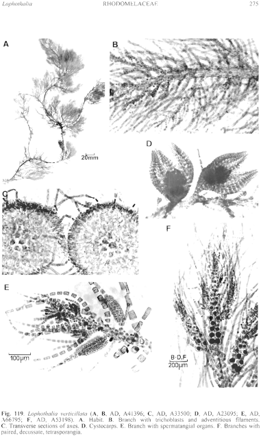
Figure 119 enlarge
Fig. 119. Lophothalia verticillata (A, B, AD, A41396; C, AD, A33500; D, AD, A23095; E, AD, A66795; F, AD, A53198). A. Habit. B. Branch with trichoblasts and adventitious filaments. C. Transverse sections of axes. D. Cystocarps. E. Branch with spermatangial organs. F. Branches with paired, decussate, tetrasporangia.
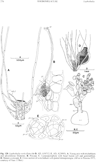
Figure 120 enlarge
Fig. 120. Lophothalia verticillata (A–D, AD, A30712; E, AD, A23095). A. Young axis with trichoblasts and adventitious filaments. B. Procarp. C. Carposporophyte with basal fusion cell and gonimoblast. D. Mature cystocarp. E. Cross section of a stichidium with paired tetrasporangia. (All as in Parsons 1975, courtesy of Aust. J. Bot.)

|
Email Contact: State Herbarium of South Australia |

|