|
|
|
|
|
|||||||||||
|
Electronic Flora of South Australia Species Fact Sheet
Phylum Rhodophyta – Order Ceramiales – Family Ceramiaceae – Tribe Griffithsieae
Selected citations: Shepherd & Womersley 1971: 166 (nomen nudum). Silva et al. 1996: 412.
Thallus (Pl. 1 fig. 3; Fig. 151C) medium to dark red, 1–3 cm high, filaments erect, subdichotomous several times, ecorticate, forming dense pulvinate masses. Attached at the base by anastomosing rhizoids; epilithic. Structure. Cells (Fig. 152G) globose near the thallus apex and 750–900 (–1200) µm in diameter, in mid and basal thallus (Fig. 153A) clavate to obovoid, 1–2 (–2.1) mm in diameter and L/D 2–3.
Reproduction: Gametophytes dioecious. Female axis 3-celled, subapical but displaced laterally by continued growth of the vegetative apical cell, flanked by a pair of hair-like synchronic laterals. Procarp systems (Fig. 152E) subapical, each with a sterile lateral cell, and supporting cell bearing a sterile cell apically and a lateral, recurved carpogonial branch of 4 cells. The inner of two connecting cells, cut off from the carpogonium after fertilisation, fuses with the supporting cell (Fig. 152E), which cuts off an apical auxiliary cell; post-fertilisation fusion cell columnar, bearing 1–3 gonimolobes terminally, most cells of which become ovoid carposporangia 25–30 µm in diameter; hypogenous cell producing abaxially 9–12 (–14) 2-celled involucral branches (Fig. 152F), of which the larger, incurved apical cells are occasionally furcate, 325–350 µm in diameter and L/D 1.8–2. Growth of the vegetative axis is often arrested during procarp development, and 1 (–4) vegetative laterals arise from the vegetative subapical cell, displacing the carposporophyte to an apparently terminal position (Fig. 152G). Spermatangia borne on numerous, minute fascicles from the subapical cell and occasionally the cell below, clustered in the constrictions between the cells; involucre absent. A fascicle is at first polychotomous, but elongation of some cells establishes an axis of 3–4 terete cells bearing 1–4 polychotomous branches, the end cells of which each produce 1–2 spermatangia.
Tetrasporangia borne in cell constrictions on numerous minute fascicles 1–3 times di- or trichotomous with small, terete cells, each of which produces a whorl of 1–6 lachrimiform tetrasporangia, 55–88 µm in diameter, or a 1-celled involucral branch (Fig. 152H) equal in size to a tetrasporangium. The sterile involucral cells stain lightly, in contrast to sporangia, which stain densely and divide when quite small.
Type from Port Elliot, S. Aust., in the upper sublittoral on granite rock (Baldock, 4.v.1963); holotype and isotypes in AD, A26365.
Selected specimens: Streaky Bay, S. Aust. (Parsons, 25.viii.1967; AD, A31933). Cape Carnot, S. Aust., mid eulittoral on walls of a shaded cavern (Womersley, 8.i.1951; AD, Al 5098). Pondalowie Bay, Yorke Pen., S. Aust., eulittoral on rocks in sheltered areas (Baldock, 14.iv.1963; AD, A26363). Robe, S. Aust., lower eulittoral ( Womersley, 30.ix.1993; AD, A63203 -"Marine Algae of southern Australia" No. 177a). Nora Creina, S. Aust., lower eulittoral (Womersley, 3.ix.1971; AD, A39547 -"Marine Algae of southern Australia" No. 177). Bridgewater Bay, Vic., upper sub littoral-lower eulittoral (Womersley, 25.i.1967; AD, A32778). Point Nepean, Vic., in rock pools (Ducker, 9.v.1965; AD, A37730).
Distribution: Streaky Bay, S. Aust. to Port Phillip Heads, Victoria.
Taxonomic notes: G. pulvinata is common in small, low, cushion-shaped masses on rock in the upper sublittoral to lower eulittoral, usually in shaded positions. Shepherd & Womersley (1971, p. 166) recorded this as a nomen nudum for Pearson I., South Australia.
G. pulvinata reproductively belongs to the G. monilis group and some intergrades with sparse branching and inflated cells resemble G. monilis and G. pilalyea. However, the erect filaments, pulvinate habit, clavate cells towards the thallus base (see Fig. 153A, B, E), and the small involucral cells of the tetrasporangial fascicles readily distinguish G. pulvinata.
References:
BALDOCK, R.N. (1976). The Griffithsieae group of the Ceramiaceae (Rhodophyta) and its southern Australian representatives. Aust. J. Bot. 24, 509–593.
SHEPHERD, S.A. & WOMERSLEY, H.B.S. (1971). Pearson Island Expedition 1969.7. The subtidal ecology of benthic algae. Trans. R. Soc. S. Aust. 95(3), 155–167.
The Marine Benthic Flora of Southern Australia Part IIIC complete list of references.
Publication:
Womersley, H.B.S. (24 December, 1998)
The Marine Benthic Flora of Southern Australia
Rhodophyta. Part IIIC. Ceramiales – Ceramiaceae, Dasyaceae
©State Herbarium of South Australia, Government of South Australia
Illustrations in Womersley Part IIIA, 1998: PLATE 1 fig. 3; FIGS 151C, 152 E–H, 153A.
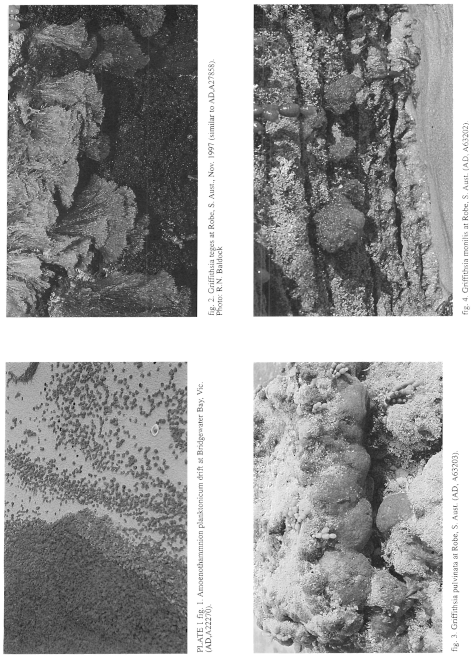
Plate 1 enlarge
PLATE 1 fig. 1. Amoenothamnion planktonium drift at Bridgewater Bay, Vic. (AD, A22270).
fig. 2. Griffithsia teges at Robe, S. Aust., Nov. 1997 (similar to Ad, A27858). Photo: R.N. Baldock.
fig. 3. Griffithsia pulvinata at Robe, S. Aust. (AD, A63203).
fig. 4. Griffithsia monilis at Robe, S. Aust. (AD, A63202)
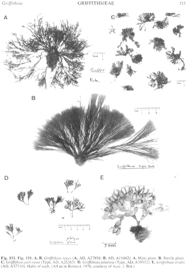
Figure 151 enlarge
Fig. 151. A, B. Griffithsia teges (A, AD, A27858; B, AD, A 11042). A. Male plant. B. Sterile plant. C. Griffithsia pulvinata (Type, AD, A26365). D. Griffithsia pilalyea (Type, AD, A39552). E. Griffithsia ovalis (AD, A37310). Habit of each. (All as in Baldock 1976, courtesy of Aust. J. Bot.)
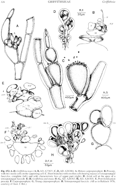
Figure 152 enlarge
Fig. 152. A–D. Griffithsia teges (A, B, AD, A27821; C, D, AD, A26386). A. Mature carposporophyte. B. Procarp, with two sterile cells on the supporting cell. C. Short branches with swollen cells bearing masses of tetrasporangial fascicles, complete (left) and with characteristic loss of upper part (right). D. Axial cell at the apex of a tetrasporangial fascicle. E–H. Griffithsia pulvinata (E–G, AD, A26363; H, AD, A26365). E. Post-fertilisation procarp. F. Carposporophyte. G. Young carposporophyte. H. Tetrasporangial fascicle. (All as in Baldock 1976, courtesy of Aust. J. Bot.)
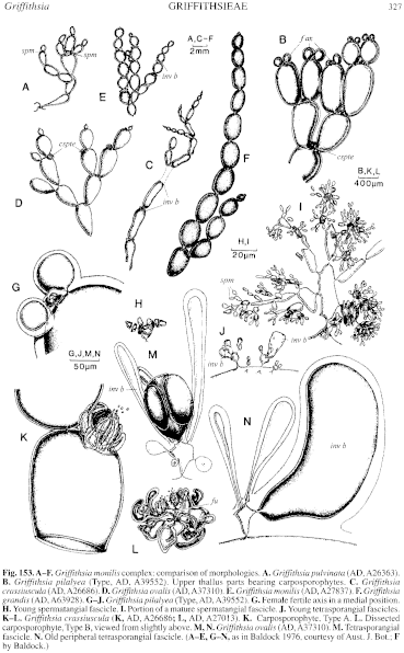
Figure 153 enlarge
Fig. 153. A–F. Griffithsia monilis complex: comparison of morphologies. A. Griffithsia pulvinata (AD, A26363). B. Griffithsia pilalyea (Type, AD, A39552). Upper thallus parts bearing carposporophytes. C. Griffithsia crassiuscula (AD, A26686). D. Griffithsia ovalis (AD, A3731.0). E. Griffithsia monilis (AD, A27837). F. Griffithsia grandis (AD, A63928). G–J. Griffithsia pilalyea (Type, AD, A39552). G. Female fertile axis in a medial position. H. Young spermatangial fascicle. I. Portion of a mature spermatangial fascicle. J. Young tetrasporangial fascicles. K–L. Griffithsia crassiuscula (K, AD, A26686; L, AD, A27013). K. Carposporophyte, Type A. L. Dissected carposporophyte, Type B, viewed from slightly above. M, N. Griffithsia ovalis (AD, A37310). M. Tetrasporangial fascicle. N. Old peripheral tetrasporangial fascicle. (A–E, G–N, as in Baldock 1976, courtesy of Aust. J. Bot.; F by Baldock.)

|
Email Contact: State Herbarium of South Australia |

|