|
|
|
|
|
|||||||||||
|
Electronic Flora of South Australia Species Fact Sheet
Phylum Rhodophyta – Class Florideophyceae – Order Corallinales – Family Corallinaceae – Subfamily Melobesioideae
Thallus normally pinkish to gray-pink, discoid to foliose, mostly 20–150 mm across and 1–3 mm thick, epigenous and usually loosely affixed, commonly with lamellate branches or perpendicularly orientated struts 1–30 mm long arising from the ventral surface, dorsal surface more or less smooth, often somewhat shiny when dried and without branching. Structure pseudoparenchymatous with dorsiventral organisation throughout; construction monomerous, consisting of a single system of branched, laterally cohering, filaments that collectively contribute to a centrally situated core and dorsal and ventral peripheral regions where portions of core filaments or their derivatives curve outwards towards the thallus surface, each filament composed of cells 5–15 µm in diameter and 5–30 µm long; epithallial cells 4–13 µm in diameter and 2–5 µm long, terminating most filaments at the thallus surface, with distal walls rounded or flattened but not flared; cell elongation occurring mainly behind actively dividing subepithallial initials that are usually as short as or shorter than their immediate inward derivatives; fusions between cells of adjacent filaments present; secondary pit-connections, haustoria, and trichocytes unknown.
Reproduction: Vegetative reproduction unknown. Gametangial thalli and carposporophytes borne in uniporate conceptacles. Tetrasporangia produced in multiporate conceptacles. Bisporangia unknown.
Gametangial thalli apparently dioecious; carpogonial filaments not seen. Mature female carposporangial conceptacle roofs protruding slightly above or flush with surrounding thallus surface, 50–75 µm thick, composed of 6–10 layers of cells above the chamber, conceptacle chambers 510–600 µm in diameter and 150–200 µm high. Mature carposporophytes apparently lacking a conspicuous central fusion cell and consisting of several-celled gonimoblast filaments bearing terminal carposporangia. Both unbranched and branched spermatangial filaments present, arising from the floor, walls and roof of male conceptacle chambers, mature male conceptacle roofs protruding slightly above or flush with surrounding thallus surface, 35–40 µm thick, composed of 5–7 layers of cells above the chamber, conceptacle chambers 220–520 µm in diameter and 50–80 µm high.
Tetrasporangial conceptacle roofs protruding above or flush with surrounding surface, 5–7 cells thick above the chamber, pore canals lined by cells that are similar in size and shape to other roof cells; conceptacle chambers 220–500 µm in diameter and 95–175 µm high, usually filled with enlarged sterile irregularly shaped vegetative cells interspersed amongst the sporangia and possessing distinct layers of darkly staining cells beneath the chamber floor, tetrasporangia scattered across the conceptacle chamber floor, each mature sporangium 30–60 µm in diameter and 70–125 µm long, containing zonately arranged tetraspores and possessing an apical plug that blocks a roof pore prior to spore release.
Type from Ninepin Point, D'Entrecasteaux Channel, Tas., 6–12 m deep (Kraft, 4.i.1993); holotype in LTB (16679); depicted in part in Wilks & Woelkerling (1994, p. 216, fig. 11A).
Selected specimens: Cape Buffon, S. Aust., 5 m deep (Kildea & Collings, 27.ix.1992; AD, A61770 = LTB, 16582). 1.3 km off Middle Point, Cape Northumberland, S. Aust., 15 m deep (Shepherd, 14.xi.1975; AD, A46708). The Gap, Cunningham Bay, Phillip I., Vic., 2 m deep (Kemp, iv.1982; LTB, 12854). Walkerville, Vic., 2–3 m deep (Platt et al., 30.xi.1983; LTB, 13912). Spike I., Bass Strait, Tas., 6 m deep (Leach, 28.ii.1979; LTB, 11601). North West Islet, Killiekranke Bay, Flinders I., Tas., 15 m deep (AIMS-NCI, Q66C 3574-Z, 21.ii.1990; AD, A60388 = LTB, 16678). Three Hummock I., NW Tas., 15 m deep (AIMS-NCI, Q66C 4952-Y, 9.ii.1991; AD A61382). Stapleton Point, Prosser Bay, Tas., 10 m deep (Olsen, 28.vii.1966; AD, A30645). Deep Glen Bay, Forestier Pen., Tas., 8–10 m deep (Gowlett-Holmes, 15.ix.1995; AD, A64476). Ninepin Point, D'Entrecasteaux Channel, Tas., 4–6 m deep (Platt & Brown, 18.ii.1983; LTB, 12760 and Platt, 18.ii.1983; LTB, 12787). Southport (Convict Burial Ground, S of Sister Bay), Tas., 3–4 m deep (Platt, 16.ii.1983; LTB, 12896). Lady Bay, Southport, Tas., 7 m deep (Brown & Kenchington, 14.x.1986; AD, A57695).
Distribution: Cape Buffon, S. Aust., to Walkerville, Vic., and around Tasmania.
Taxonomic notes: Phymatolithon masonianum has been found at depths of 2–15 m on sponges and on rock. Thalli of P. masonianum are usually easily recognised in the field from their distinctive discoid to foliose growth form and large thalli with smooth dorsal surfaces that often look somewhat shiny, with struts on the ventral surface; no other known southern Australian non-geniculate coralline shows this combination of features. Only one dried male thallus and two dried carposporangial thalli are known; carpogonial filaments have yet to be found, meaningful measurements of carposporangia could not be obtained, and the ontogeny of all reproductive structures requires further investigation.
References:
WILKS, K.M. & WOELKERLING, W.J. (1994). An account of southern Australian species of Phymatolithon (Corallinaceae, Rhodophyta) with comments on Leptophytum. Aust. Syst. Bot. 7, 183–223.
The Marine Benthic Flora of Southern Australia Part IIIB complete list of references.
Publication:
Womersley, H.B.S. (28 June, 1996)
The Marine Benthic Flora of Southern Australia
Rhodophyta. Part IIIB. Gracilarialse, Rhodymeniales, Corallinales and Bonnemaisoniales
Reproduced with permission from The Marine Benthic Flora of Southern Australia Part IIIB 1996, by H.B.S. Womersley. Australian Biological Resources Study, Canberra. Copyright Commonwealth of Australia.
Illustrations in Womersley Part IIIA, 1996: PLATE 3 fig. 2; FIGS 63A, 78, 79.
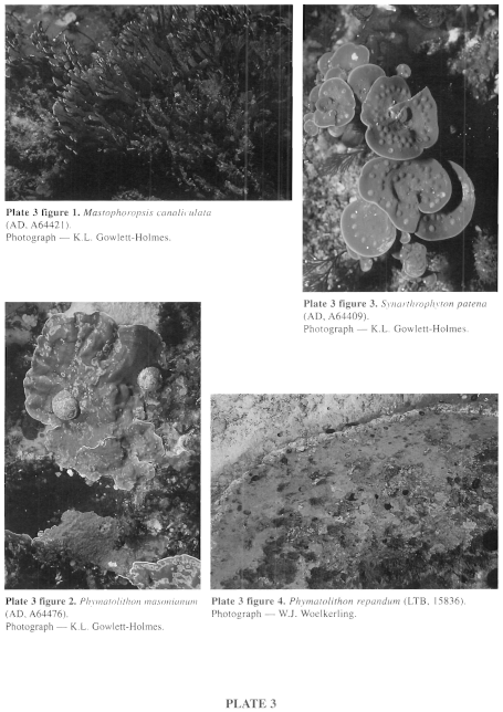
Plate 3 enlarge
PLATE 3
figure 1. Mastophoropsis canaliculata (AD, 64421). Photograph K.L. Gowlett-Holmes.
figure 2. Phymatolithon masonianum (AD, A64476). Photograph - K.L.Gowlett-Holmes.
figure 3. Synarthrophyton patena (AD, A64409). Photograph - K.L.Gowlett-Holmes.
figure 4. Phymatolithon repandum (LTB, 15836). Photograph - W.J. Woelkerling.
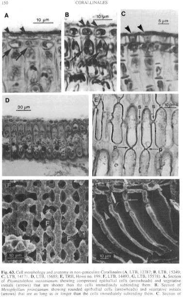
Figure 63 enlarge
Fig. 63. Cell morphology and anatomy in non-geniculate Corallinales (A, LTB, 12787; B, LTB, 15249; C, LTB, 14171; D, LTB, 15688; E, TRH, Howe no. 199; F, LTB, 14493; G, LTB, 15578). A. Section of Phymatolithon masonianum showing compressed epithallial cells (arrowheads) and vegetative initials (arrows) that are shorter than the cells immediately subtending them. B. Section of Mesophyllum printzianum showing rounded epithallial cells (arrowheads) and vegetative initials (arrows) that are as long as or longer than the cells immediately subtending them. C. Section of Sporolithon durum showing epithallial cells (arrow heads) with flared outer walls D. Palisade cells of contiguous filaments of Metamastophora flabellata showing cell-fusions (F). E. Columnar cells of Lithophyllum frondosum showing primary (arrow - p) and secondary (arrow - s) pit connections. F. Fracture of Sporolithon durum showing fusions (F) between cells of contiguous filaments. G. Fracture of Lithophyllum corallinae showing pit connections in surface (arrowhead) and sectional (arrow) views.
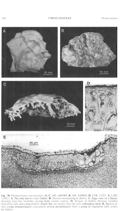
Figure 78 enlarge
Fig. 78. Phymatolithon masonianum (A, C, AD, A60388; B, AD, A30645; D, LTB, 12787; E, LTB, 11601). A. The only known male thallus. B. Discoid tetrasporangial thallus. C. Edge view of a thallus showing strut-like branches arising from ventral surface. D. Section of thallus showing rounded epithallial cells and subepithallial initials that are shorter than the cells subtending them. E. Section of very young tetrasporangial conceptacle arising adventitiously from a group of vegetative cells within the thallus.
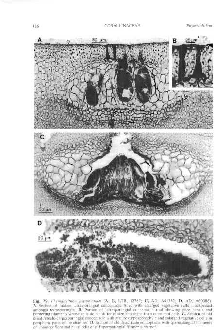
Figure 79 enlarge
Fig. 79. Phymatolithon masonianum (A, B, LTB, 12787; C, AD, A61382; D, AD, A60388). A. Section of mature tetrasporangial conceptacle filled with enlarged vegetative cells interspersed amongst tetrasporangia. B. Portion of tetrasporangial conceptacle roof showing pore canals and bordering filaments whose cells do not differ in size and shape from other roof cells. C. Section of old dried female-carposporangial conceptacle with mature carposporophyte and enlarged vegetative cells in peripheral parts of the chamber. D. Section of old dried male conceptacle with spermatangial filaments on chamber floor and basal cells of old spermatangial filaments on roof.

|
Email Contact: State Herbarium of South Australia |

|