|
|
|
|
|
|||||||||||
|
Electronic Flora of South Australia Species Fact Sheet
Phylum Rhodophyta – Class Florideophyceae – Order Rhodymeniales – Family Champiaceae
Thallus (Fig. 54D) red to red-brown, erect and spreading, 3–11 cm high, much branched with one to several axes irregularly radially branched for 3 or 4 orders. Axes and branches terete, 1–1.5 mm in diameter below, tapering gradually to branchlets 0.3–0.5 mm in diameter, young branches slightly constricted at diaphragms, segments L/D 1–1.5, ends straight, apices rounded; diaphragms distinct throughout most of thallus, obscured near older bases. Holdfast small, discoid, with occasional haustorial pads; epiphytic on Amphibolis. Structure multiaxial, with 10–15 apical cells, developing a cortex of angular cells 25–50 (–60) µm across and mostly L/B (1.5–) 2–3, with small cells cut off from their corners and forming a continuous outer cortex (to 3 cells thick) on older axes (Fig. 53J); longitudinal filaments (Fig. 53H) peripheral only, usually with two (–3) complete cells (with secretory cells) and two part cells between the diaphragms (Fig. 531). Rhodoplasts discoid, usually in chains.
Reproduction: Gametangial thalli dioecious. Carpogonial branches 4-celled, borne on a cortical (supporting) cell together with a 2-celled auxiliary cell branch (Fig. 53K). Carposporophyte (Fig. 53L) with a basal fusion cell, branched gonimoblast filaments of oyoid cells and terminal ovoid to obconical carposporangia (40–) 50–90 µm in diameter. Basal nutritive tissue slight, inner cells of pericarp stellate and separating. Cystocarps (Fig. 54E) scattered, subspherical to urceolate, 0.7–1.0 (–1.2) mm in diameter, pericarp 60–130 µm and 3–5 cells thick, ostiolate. Spermatangia covering segments near apices of young branches, cut off from branched filaments of initials (Fig. 53M), ovoid, 3–5 µm in diameter.
Tetrasporangia (Fig. 54F) scattered, transformed from cortical cells, (50–) 60–120 µm in diameter, tetrahedrally divided (Fig. 53N).
Type (of variety) from Tiparra Reef, Spencer Gulf, S. Aust., 11 m deep (Shepherd, 23.xii.1970); holotype in AD, A38255.
Selected specimens: The type and Tiparra Reef, S. Aust., 5 m deep (Shepherd, 30.ix.1970; AD, A37291) and 11 m deep (Shepherd, 13.xii.1971; AD, A41276).
Distribution: The var. amphibolis only known from Tiparra Reef, S. Aust.
Taxonomic notes: The Tiparra Reef plants were considered distinct as a variety by Reedman & Womersley, characterised by dense radial branching, size and being epiphytic on Amphibolis.
References:
REEDMAN, D.J. & WOMERSLEY, H.B.S. (1976). Southern Australian species of Champia and Chylocladia (Rhodyméniales: Rhodophyta). Trans. R. Soc. S. Aust. 100, 75–104.
The Marine Benthic Flora of Southern Australia Part IIIB complete list of references.
Publication:
Womersley, H.B.S. (28 June, 1996)
The Marine Benthic Flora of Southern Australia
Rhodophyta. Part IIIB. Gracilarialse, Rhodymeniales, Corallinales and Bonnemaisoniales
Reproduced with permission from The Marine Benthic Flora of Southern Australia Part IIIB 1996, by H.B.S. Womersley. Australian Biological Resources Study, Canberra. Copyright Commonwealth of Australia.
Illustrations in Womersley Part IIIA, 1996: FIGS 53 H–N, 54 D–F.
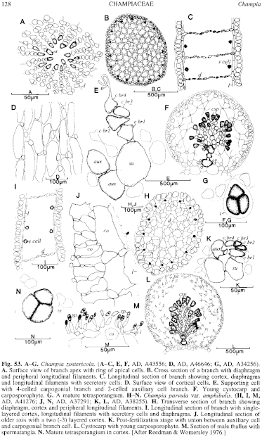
Figure 53 enlarge
Fig. 53. A–G. Champia zostericola. (A–C, E, F, AD, A43556; D, AD, A46646; G, AD, A34256). A. Surface view of branch apex with ring of apical cells. B. Cross section of a branch with diaphragm and peripheral longitudinal filaments. C. Longitudinal section of branch showing cortex, diaphragms and longitudinal filaments with secretory cells. D. Surface view of cortical cells. E. Supporting cell with 4-celled carpogonial branch and 2-celled auxiliary cell branch. F. Young cystocarp and carposporophyte. G. A mature tetrasporangium. H–N. Champia parvula var. amphibolis. (H, I, M, AD, A41276; J, N, AD, A37291; K, L, AD, A38255). H. Transverse section of branch showing diaphragm, cortex and peripheral longitudinal filaments. I. Longitudinal section of branch with single-layered cortex, longitudinal filaments with secretory cells and diaphragms. J. Longitudinal section of older axis with a two (-3) layered cortex. K. Post-fertilization stage with union between auxiliary cell and carpogonial branch cell. L. Cystocarp with young carposporophyte. M. Section of male thallus with spermatangia. N. Mature tetrasporangium in cortex. [After Reedman & Womersley 1976.]
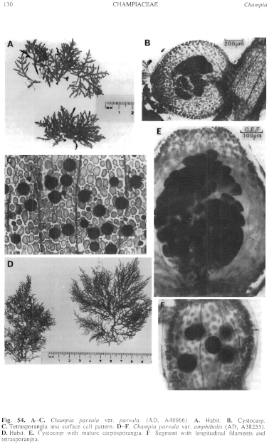
Figure 54 enlarge
Fig. 54. A–C. Champia parvula var. parvula. (AD, A48966). A. Habit. B. Cystocarp. C. Tetrasporangia and surface cell pattern. D–F. Champia parvula var. amphibolis (AD, A38255). D. Habit. E. Cystocarp with mature carposporangia. F. Segment with longitudinal filaments and tetrasporangia.

|
Email Contact: State Herbarium of South Australia |

|