|
|
|
|
|
|||||||||||
|
Electronic Flora of South Australia Species Fact Sheet
Phylum Rhodophyta – Class Florideophyceae – Order Bonnemaisoniales – Family Bonnemaisoniaceae
Selected citations: May 1965: 374.
Synonym
Bonnemaisonia asparagoides (Woodward) C. Agardh var. hypnoides Reinbold 1899: 47. Lucas 1929b: 50. Lucas & Perrin 1947: 243. Tisdall 1898: 512. Wilson 1892: 169. Womersley 1950: 163.
Thallus (Fig. 149A) medium red, 4–12 cm high, delicate, much branched and spreading, branching essentially distichous on long main laterals, with main branches terete, 400–600 (–800) µm broad, bearing opposite simple branchlets, alternately long and short; the longer 1.5–2.5 (–3.5) mm long and 100–150 (–200) µm in basal diameter and tapering gently, the lower ones with hamate ends, and the opposite short branchlets often becoming fertile or developing as longer branches. Attachment mainly by lower hamate branchlets; probably epiphytic. Structure uniaxial (Fig. 149B), with a small but prominent apical cell, the subapical cells cutting off two opposite periaxial cells which develop alternately into full sized branchlets and opposite short branchlets; periaxial cells dividing, mainly basally, to produce 2–4 primary cortical cells which cover the axial cell which becomes 15–30 µm in diameter and L/D (10–) 20–30; cortex separated from the axial filament by a space, mature primary cortical cells subspherical, 30–45 µm in diameter, with small secondary cortical cells forming rosettes, often bearing slender hairs. Branchlets (Fig. 149B) with 3 periaxial cells, becoming radially elongate and each with 4 primary cortical cells becoming subspherical and 20–28 µm in diameter, with sparse small outer cells. Rhodoplasts ribbon shaped, numerous per cell.
Reproduction: Gametangial thalli monoecious (Fig. 149D), reproductive organs formed on the shorter of the branchlet pairs. Carpogonial branches 3-celled, with a lateral tuft of nutritive filaments, borne on a stalk 5–6 axial cells long. Cystocarps (Fig. 149E) pedicellate, broadly urceolate and ostiolate, 400–500 µm in diameter, gonimoblast much branched, arising from a basal fusion cell, with large, terminal, elongate-ovoid carposporangia 30–50 µm in diameter; pericarp of numerous, branched, erect filaments with an outer cortical layer 1–2 cells thick. Spermatangial organs (Fig. 149D) with a firm mucilaginous sheath, ovoid, 100–130 µm long and 60–90 µm in diameter, with a row of 4–6 axial cells each cutting off several periaxial cells which produce clusters of initials which cut off ovoid spermatangia about 2 µm in diameter.
Tetrasporophyte unknown.
Type from Port Phillip Heads, Vic. (Wilson, 1884); holotype in Herb. Agardh, LD, 37379.
Selected specimens: Whyalla, S. Aust., 12 m deep (Branden, 15.v.1984; AD, A55402). Fitzgerald Bay, Point Lowly, S. Aust., 14 m deep (Branden, 13.ix.1987; AD, A59340). Blanche Harbor, N Spencer Gulf, S. Aust., 13 m deep (Branden, 10.ix.1987; AD, A59351). N Spencer Gulf, S. Aust., 10 m deep (Shepherd, 5.ix.1973; AD, A44233). Sturt Bay, S. Aust., drift (Davey; AD, A442). 10 km W of Outer Harbor, S. Aust., 23 m deep (R. Lewis, 10.ix.1972; AD, A42659). Brighton, S. Aust., drift (Bienert, 12.xi.1965; AD, A29661). Pennington Bay, Kangaroo I., S. Aust., drift (Womersley, 29.viii.1948; AD, A8744). Low Head, Tas., 2–12 m deep (Perrin, Oct. 1937; AD, A8500).
Distribution: Whyalla to Brighton, S. Aust., Port Phillip, Vic., and Low Head, Tasmania.
Taxonomic notes: B. australis is usually a deep water alga, most known specimens being from the Gulf region of South Australia.
It differs from B. asparagoides of European coasts in being more lightly corticated, with periaxial filaments less branched (2–3 branches before the cortex in the latter), in having slenderer axial filaments (15–30 µm compared to 40–60 µm), in that the small outer, rounded, cortical cells do not appear as gland cells, and in having numerous lower branchlets with hamate ends.
The Sturt Bay (Davey) specimen (AD, A442) is probably an isotype of B. asparagoides var. hypnoides Reinbold.
References:
LEVRING, T. (1953). The marine algae of Australia. I. Rhodophyta: Goniotrichales, Bangiales and Nemalionales. Arkiv för Bot. Ser. 2, 2, 457–530.
LUCAS, A.H.S. & PERRIN, F. (1947). The Seaweeds of South Australia. Part 2. The Red Seaweeds. (Govt Printer: Adelaide.)
LUCAS, A.H.S. (1929b). A census of the marine algae of South Australia. Trans. R. Soc. S. Aust. 53, 45–53.
MAY, V. (1965). A census and key to the species of Rhodophyceae (red algae) recorded from Australia. Contr. N.S.W. natn. Herb. 3, 349–429.
REINBOLD, T. (1899). Meeresalgen von Investigator Street (Slid Australien), gesammelt von Miss Nellie Davey (Waltham, Honiton). Hedwigia 38, 39–51.
TISDALL, H.T. (1898). The algae of Victoria. Rep. 7th Meet. Aust. Ass. Adv. Sci., Sydney, 1898, pp. 493–516.
WILSON, J.B. (1892). Catalogue of algae collected at or near Port Phillip Heads and Western Port. Proc. R. Soc. Vict. 4, 157–190.
WOMERSLEY, H.B.S. (1950). The marine algae of Kangaroo Island. III. List of Species 1. Trans. R. Soc. S. Aust. 73, 137–197.
The Marine Benthic Flora of Southern Australia Part IIIB complete list of references.
Publication:
Womersley, H.B.S. (28 June, 1996)
The Marine Benthic Flora of Southern Australia
Rhodophyta. Part IIIB. Gracilarialse, Rhodymeniales, Corallinales and Bonnemaisoniales
Reproduced with permission from The Marine Benthic Flora of Southern Australia Part IIIB 1996, by H.B.S. Womersley. Australian Biological Resources Study, Canberra. Copyright Commonwealth of Australia.
Illustration in Womersley Part IIIA, 1996: FIG. 149.
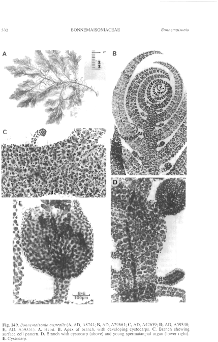
Figure 149 enlarge
Fig. 149. Bonnemaisonia australis (A, AD, A8744; B, AD, A29661; C, AD, A42659; D, AD, A59340; E, AD, A59351). A. Habit. B. Apex of branch, with developing cystocarps. C. Branch showing surface cell pattern. D. Branch with cystocarp (above) and young spermatangial organ (lower right). E. Cystocarp.

|
Email Contact: State Herbarium of South Australia |

|