|
|
|
|
|
|||||||||||
|
Electronic Flora of South Australia Species Fact Sheet
Phylum Rhodophyta – Class Florideophyceae – Order Nemaliales – Family Galaxauraceae
Selected citations: Millar 1990: 307, fig. 7A–C.
Thallus (Fig. 27D) medium grey-red to dark red, sometimes brownish, 5–20 (–38) cm high, subdichotomously branched every 1–3 (–11) cm usually with constrictions at points of branching, segments 1.5–3 mm in diameter below, 3–4 mm in diameter above, ultimate segments tapering to a point. Holdfast discoid, 1.5–2 mm across; epilithic. Structure multiaxial, with a central core of slender, entwined filaments 2–3 (–6) µm in diameter, from which radiate medullary filaments giving rise to a cortex (Fig. 27E) of 1–2 layers of ovoid, rhodoplastic, cells 10–15 µm in diameter and an outer layer of colourless utricles, polygonal in surface view, 25–40 µm long and 18–25 (–30) µm in diameter.
Reproduction: Sexual thalli monoecious. Carpogonial branches (Fig. 28C) 3-celled, developed on outer medullary cells near the apices, with the hypogynous cell producing four sterile branches, usually a 3-celled branch, two 2-celled branches and a 1-celled branch, and the basal cell producing several lateral branches which form the involucre after fertilization. Fertilized carpogonium producing 3–4 initials with the gonimoblast filaments (Fig. 28D) developing chains of ovoid to pyriform carposporangia 15–20 µm long and 5–7 µm in diameter; fusion cell present. Cystocarps (Fig. 27F) 200–250 µm in diameter, ostiolate, with a well developed involucre. Spermatangial branches (Fig. 27E) arising from hypodermal cortical cells and projecting between surface utricles, 2–3 cells long, producing 2–3 spermatangial initials each with 1–3 ovoid spermatangia 2–3 µm in diameter.
Type from Ocean Reef Marina, Sorrento, W. Aust. (O'Brien & Millar, 9.xii.1980); holotype in MELU, 24309.
Selected specimens: (see also Huisman 1986, p. 279): Point Clune, Rottnest I., W. Aust., 13–15 m deep (Kraft & Ricker, 5.xii.1980; MELU, 24308). Point Sinclair, S. Aust., drift (Humphrey, Feb. 1954; AD, A56759). Daly Head, Yorke Pen., S. Aust., drift (Gordon, 26.iii.1967; AD, A31579). Marino, S. Aust., drift (Womersley, 21.v.1953; AD, A18633). Port Elliot, S. Aust., drift (Kraft 3518, 30.iv.1971; AD, A45969). Warrnambool, Vic., drift (Kraft, 11.ii.1984; MELU, 24357). Aireys Inlet, Vic., drift (Kraft, 21.ii.1976; MELU, 23231). Collaroy, N.S.W., drift (May 2028, 22.i.1946; NSW). Korffs I., Coffs Harbour, N.S.W., 20 m deep (Millar & Gabrielson, 22.i.1982; MELU, AM9I 8).
Distribution: Greenough Point (10 km S of Geraldton), W. Aust., around southern Australia (not Tasmania) to Coffs Harbour, N.S.W.; Norfolk I.
References:
HUISMAN, J.M. (1986). The red algal genus Scinaia (Galaxauraceae, Nemaliales) from Australia. Phycologia 25, 271–296.
MILLAR, A.J.K. (1990). Marine Red Algae of the Coffs Harbour Region, northern New South Wales. Aust. Syst. Bot. 3, 293–593.
The Marine Benthic Flora of Southern Australia Part IIIA complete list of references.
Publication:
Womersley, H.B.S. (14 January, 1994)
The Marine Benthic Flora of Southern Australia
Rhodophyta. Part IIIA, Bangiophyceae and Florideophyceae (to Gigartinales)
Reproduced with permission from The Marine Benthic Flora of Southern Australia Part IIIA 1994, by H.B.S. Womersley. Australian Biological Resources Study, Canberra. Copyright Commonwealth of Australia.
Illustrations in Womersley Part IIIA, 1994: FIGS 27 D–F, 28C, D.
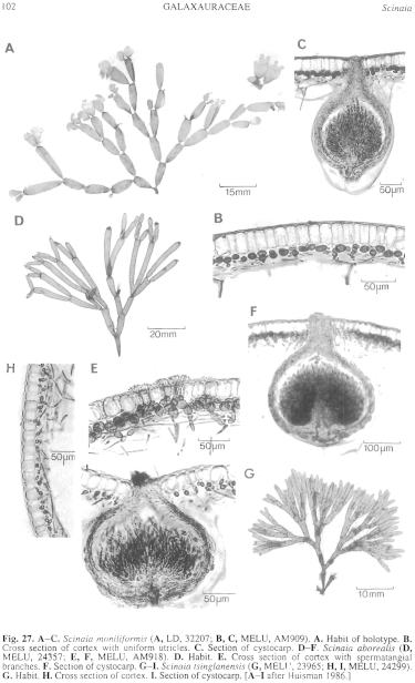
Figure 27 enlarge
Fig. 27. A–C. Scinaia moniliformis (A, LD, 32207; B, C, MELU, AM909). A. Habit of holotype. B. Cross section of cortex with uniform utricles. C. Section of cystocarp. D–F. Scinaia aborealis (D, MELU, 24357; E, F, MELU, AM918). D. Habit. E. Cross section of cortex with spermatangial branches. F. Section of cystocarp. G–I. Scinaia tsinglanensis (G, MELU, 23965; H, I, MELU, 24299). G. Habit. H. Cross section of cortex. I. Section of cystocarp. [A–I after Huisman 1986.]
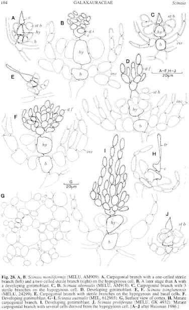
Figure 28 enlarge
Fig. 28. A, B. Scinaia moniliformis (MELU, AM909). A. Carpogonial branch with a one-celled sterile branch (left) and a two-celled sterile branch (right) on the hypogynous cell. B. A later stage than A with a developing gonimoblast. C, D. Scinaia aborealis (MELU, AM918). C. Carpogonial branch with 3 sterile branches on the hypogynous cell. D. Developing gonimoblast. E, F. Scinaia tsinglanensis (MELU, 24299). E. Carpogonial branch with sterile branches on the hypogynous and basal cells. F. Developing gonimoblast. G–I. Scinaia australis (MEL, 612903). G. Surface view of cortex. H. Mature carpogonial branch. I. Developing gonimoblast. J. Scinaia proliferata (MELU, GK 4932). Mature carpogonial branch with several cells derived from the hypogynous cell. [A–J after Huisman 1986.]

|
Email Contact: State Herbarium of South Australia |

|