|
|
|
|
|
|||||||||||
|
Electronic Flora of South Australia Species Fact Sheet
Phylum Phaeophyta – Order Chordariales – Family Spermatochnaceae
Selected citations: Hamel 1937: 184, fig. 40 (v, vi, viii). Harvey 1846: p1.70. Kylin 1933: 66, figs 31, 32; 1940: 51, fig. 29. Novaczek et al. 1986: 407, figs 1–19. Oltmanns 1922: 39, fig. 334(1,2). Peters & Müller 1986c: 417, figs 1–14. Womersley 1967: 236.
Thallus (Fig. 39A) medium to dark brown, slightly mucoid, slender and much branched in an irregularly subdichotomous manner, 5–12(20) cm long with cylindrical, tapering branches 0.5–1 mm in diameter below, (100–) 150–200 µm in diameter near the apices, attached by a small discoid holdfast, usually epiphytic. Medulla (Fig. 40B) of 4–5 longitudinal filaments which separate laterally leaving a hollow medulla, filaments 4–8 (–10) in diameter with cells becoming very long. Cortex (Fig. 40C,E) 100–200 µm and 3 (–4) cells thick, with large inner cells (producing occasional hyphae) grading to the smooth-surfaced outer layer of cells which are angular in surface view (Fig. 40D), 10–15 (–20) µm broad and L/B (1–) 1.5–2.5 in lesser branches, becoming 20–40 µm broad and L/B 3–4 in older parts. Primary assimilatory filaments (Fig. 40A,B) present from near the branch apices, with associated hairs, becoming separated shortly below the apices by enlargement of cortical cells, each 3–5 cells long, usually curved, clavate, with the terminal cell subspherical to ovoid, 8–12 µm in diameter and L/B 1–1.5 (–2), with secondary assimilatory filaments (Fig. 40E) developing from adjacent outer cortical cells to give tufts of filaments 80–120 µm and 6–10 cells long, terminal cell 12–16 µm in diameter and L/B 1–1.5, the upper cells with abundant physodes, and accompanied by hairs and sporangia. Phaeoplasts (Fig. 40D) in outer cortical cells numerous, discoid, each with a pyrenoid, and with small physodes present. Phaeophycean hairs (Fig. 40B,E) present close to apices and in secondary assimilatory tufts, 10–14 µm in diameter. Growth by 4–5 apical filaments (Fig. 40A) in relatively broad apices, with the primary assimilatory filaments obscuring the apical cells.
Reproduction: Reproduction by plurilocular and unilocular sporangia borne in the secondary tufts, often together. Plurilocular sporangia (Fig. 40E) linear, uniseriate, (20–) 25–40 µm long with (4–) 6–10 locules, 4–6 µm in diameter. Unilocular sporangia (Fig. 40E) broadly clavate, sessile, 30–40 (–50) µm long and 15–2512m in diameter.
Type from England; in BM (ex K)-lectotype to be selected.
Selected specimens: Northern Spencer Gulf, S. Aust., 0–1 m deep (Shepherd, 4.ix.1973; ADU, A44097). Aldinga, S. Aust., on Cystoseira in upper sublittoral pools (Skinner, 26.x.1977; ADU, A48718). American R. inlet, Kangaroo I., S. Aust., on Cystoseira, upper sublittoral in channel (Womersley, 30.x.1966; ADU, A30889). Walkerville, Vic., drift (Sinkora A2166, 7.iii.1975; ADU, A48295).
Distribution: Europe and the N. Atlantic; Japan.
In southern Australia, from northern Spencer Gulf, S. Aust. to Walkerville, Vic. and Tasmania (Harvey 1859b, p.291), in shallow, calm water areas, usually epiphytic.
Taxonomic notes: As in Britain and Europe, Stilophora rhizodes is a shallow water species, in contrast to Spermatochnus paradoxus which is apparently only found in deep water.
References:
AGARDH, J.G. (1841). In historiam algarum symbolae auctore. Linnaea 15, 1–50, 443–457.
HAMEL, G. (1937). Phéophycées de France. Fasc. III, pp. 177–240. (Paris.)
HARVEY, W.H. (1846). Phycologia Britannica. Plates 1–72. (Reeve: London.)
HARVEY, W.H. (1859b). Algae. In Hooker, J.D., The Botany of the Antarctic Voyage. Part III. Flora Tasmaniae. Vol. 2, pp. 282–343, Plates 185–196.
KYLIN, H. (1933). Über die Entwicklungsgeschichte der Phaeophyceen. Acta Univ. lund. N.F. Avd. 2, 29(7), 1–102, Plates 1, 2.
KYLIN, H. (1940). Die Phaeophyceenordnung Chordariales. Acta Univ. lund. N.F. Avd. 2, 36(9), 1–67, Plates 1–8.
NOVACZEK, I., BIRD, C.J. & McLACHLAN, J.L. (1986). Culture and field studies of Stilophora rhizodes (Phaeophyceae, Chordariales) from Nova Scotia, Canada. Br. phycol. J. 21, 407–416.
OLTMANNS, F. (1922). Morphologie and Biologie der Algen. Vol. 2. (Jena.)
PETERS, A.F. & MÜLLER, D.G. (1986c). Sexual reproduction of Stilophora rhizodes (Phaeophyceae, Chordariales) in culture. Br. phycol. J. 21, 417–423.
WOMERSLEY, H.B.S. (1967). A critical survey of the marine algae of southern Australia. II. Phaeophyta. Aust. J. Bot. 15, 189–270.
The Marine Benthic Flora of Southern Australia Part II complete list of references.
Publication:
Womersley, H.B.S. (14 December, 1987)
The Marine Benthic Flora of Southern Australia
Part II
©Board of the Botanic Gardens and State Herbarium, Government of South Australia
Illustrations in Womersley Part II, 1997: FIGS 39A, 40 A–E.
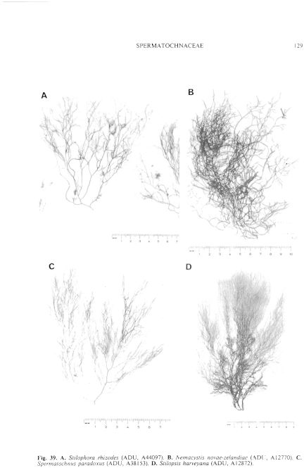
Figure 39 enlarge
Fig. 39. A. Stilophora rhizodes (ADU, A44097). B. Nemacystis novae-zelandiae (ADU, Al2770). C. Spermatochnus paradoxus (ADU, A38153). D. Stilopsis harveyana (ADU, Al2872).
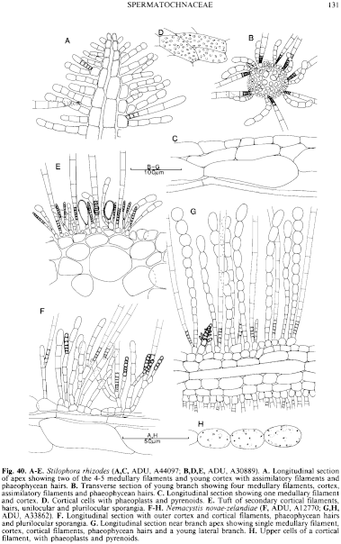
Figure 40 enlarge
Fig. 40. A–E. Stilophora rhizodes (A,C, ADU, A44097; B,D,E, ADU, A30889). A. Longitudinal section of apex showing two of the 4-5 medullary filaments and young cortex with assimilatory filaments and phaeophycean hairs. B. Transverse section of young branch showing four medullary filaments, cortex, assimilatory filaments and phaeophycean hairs. C. Longitudinal section showing one medullary filament and cortex. D. Cortical cells with phaeoplasts and pyrenoids. E. Tuft of secondary cortical filaments, hairs, unilocular and plurilocular sporangia. F–H. Nemacystis novae-zelandiae ( F, ADU, Al2770; G,H, ADU, A33862). F. Longitudinal section with outer cortex and cortical filaments, phaeophycean hairs and plurilocular sporangia. G. Longitudinal section near branch apex showing single medullary filament, cortex, cortical filaments, phaeophycean hairs and a young lateral branch. H. Upper cells of a cortical filament, with phaeoplasts and pyrenoids.

|
Email Contact: State Herbarium of South Australia |

|