|
|
|
|
|
|||||||||||
|
Electronic Flora of South Australia Species Fact Sheet
Phylum Phaeophyta – Order Chordariales – Family Scytothamnaceae
Selected citations: Clayton 1986: 371, figs 1–5, 7–10, 35, 38–41. Gibson 1908: 137, pls 490–491. Levring 1941: 627, fig. 9. Lindauer et al. 1961: 268, pl. 3. Naylor 1956: 386, figs 1,2. Nelson & Adams 1983: 87, fig. 11.
Synonym
Price 1969: 37, figs 1,2. Womersley 1967: 246.
Thallus (Pl. 3 fig. 2; Fig. 42A) dark brown, much branched, relatively cartilaginous and not mucoid, with one to many main axes each bearing 2–3 orders of irregularly arranged, terete to compressed, branches with older branches often becoming hollow, (5–) 10–30 (–40) cm long with axes 1–2 (–4) mm across and lesser branches 0.5–1 mm in diameter, tapering to both ends; axes often denuded below; epilithic, attached by a discoid to robust holdfast 1–5(10) mm across. Medulla multiaxial from a group of apical cells (Fig. 43A), producing haplostichous longitudinal filaments which become reticulate by lateral anastomoses, and differentiate into an outer medulla (Fig. 43C) of closely appressed longitudinal filaments and an inner medulla of more loosely arranged filaments 4–8 µm in diameter (cells L/B 3–10) with interwoven hyphae 5–8 µm in diameter with very long cells; medulla of axes and older branches solid and compact, with numerous hyphae. Cortex (Fig. 43B,C) dense, 40–60 µm thick, of radial rows of 2–6 more or less isodiametric cells (L/B 1–2), 6–12 µm in diameter, branched and fairly closely associated laterally, becoming pseudoparenchymatous on older branches. Phaeoplasts (Fig. 43D) stellate or lobed, with a central pyrenoid, separating into several discoid to elongate ones in some cells; physodes dense, obscuring phaeoplasts. Phaeophycean hairs grouped in shallow to deep cortical cryptostomata, 6–8 Aim in diameter.
Microthallus filamentous, branched.
Reproduction: Reproduction by ovoid to globose unilocular meiosporangia (Fig. 43B,C) lying within the cortex and visible in surface view, formed from young cortical cells and reaching to the thallus surface, 40–60 µm long and 40–50 µ in diameter. Microthallus gametophytic, filamentous, with plurilocular gametangia 28–36 µm long and 14–16 µm in diameter (Naylor 1956; Clayton 1986).
Type from New Zealand (Richard); in Herb. Agardh, LD, 46600.
Selected specimens: Apollo Bay, Vic., lower eulittoral pools ( Womersley, 23.i.1967; ADU, A31756-"Marine Algae of southern Australia" No.52). Three Hummocks I., Tas., upper sublittoral on granite ( Wollaston & Mitchell, 21.ii.1964; ADU, A27633). Boat Harbour Beach, Tas., mid eulittoral ( Womersley, 4.x.1986; ADU, A57267). Devonport, Tas., drift (Gordon, 18.i.1966; ADU, A29994). Guerilla Bay, 14 km S. of Batemans Bay, N.S.W., lower eulittoral (Barrett, 2.viii.1977; ADU, A48213). Tamarama Bay, Sydney, N.S.W., mid eulittoral (King, 28.iii.1984; ADU, A55293). Curl Curl, Sydney, N.S.W., on rocks (May 1159, 1.xii.1945; NSW).
Distribution: New Zealand, Juan Fernandez I., Chile.
In southern Australia, from Apollo Bay, Vic. to Curl Curl, Sydney, N.S.W. and around Tasmania. Lord Howe I. (in NSW).
Taxonomic notes: Scytothamnus australis is common on Tasmanian and to a lesser extent on Victorian coasts, usually in the lower to mid eulittoral, and is best developed in summer months though usually present throughout the year. It differs from S. fasciculatus in being a considerably more robust plant, with a more extensive cortex.
References:
CLAYTON, M.N. (1986). Culture studies on the life history of Scytothamnus australis and Scytothamnus fasciculatus (Phaeophyta) with electron microscope observations on sporogenesis and gametogenesis. Br. phycol. J. 21, 371–386.
GIBSON, C.M. (1908). The morphology and systematic position of Scytothamnus australis. London J. Bot. 46, 137–141, Plates 490–491.
HOOKER, J.D. & HARVEY, W.H. (1845). Algae Novae Zelandiae. Lond. J. Bot. 4, 521–551.
LEVRING, T. (1941). Die Meeresalgen der Juan Fernandez-Inseln. Nat. Hist. Juan Fernandez & Easter Is., Vol. 2, 601–670, Plates 49–53.
LINDAUER, V.W., CHAPMAN, V.J. & AIKEN, M. (1961). The marine algae of New Zealand. II. Phaeophyceae. Nova Hedwigia 3, 129–350, Plates 57–97.
NAYLOR, M. (1956). A note on the life history of Scytothamnus australis H. & H. New Phytol. 55, 386–388.
NELSON, W.A. & ADAMS, N.M. (1983). A taxonomic revision of the families Chordariaceae and Chordariopsidaceae (Phaeophyta) in New Zealand. N.Z. J. Bot. 21, 77–92.
PRICE, I.R. (1969). The structure and classification of Scytothamnus australis (J. Agardh) J.D. Hooker et Harvey (Phaeophyta). Phycologia 8, 37–41.
WOMERSLEY, H.B.S. (1967). A critical survey of the marine algae of southern Australia. II. Phaeophyta. Aust. J. Bot. 15, 189–270.
The Marine Benthic Flora of Southern Australia Part II complete list of references.
Publication:
Womersley, H.B.S. (14 December, 1987)
The Marine Benthic Flora of Southern Australia
Part II
©Board of the Botanic Gardens and State Herbarium, Government of South Australia
Illustrations in Womersley Part II, 1997: PLATE 3 fig. 2; FIGS 42A, 43 A–D.

Plate 3 enlarge
PLATE 3 fig. 1. Suringariella harveyana, with bases sand covered, Point Avoid, S. Aust. (ADU, A57368).
fig. 2. Scytothamnus australis in the mid eulittoral at Boat Harbour Beach, Tas. (ADU, A57267).
fig. 3. Scytosiphon lomentaria crusts in the lower eulittoral at Boat Harbour Beach, Tas. (ADU, A57269).
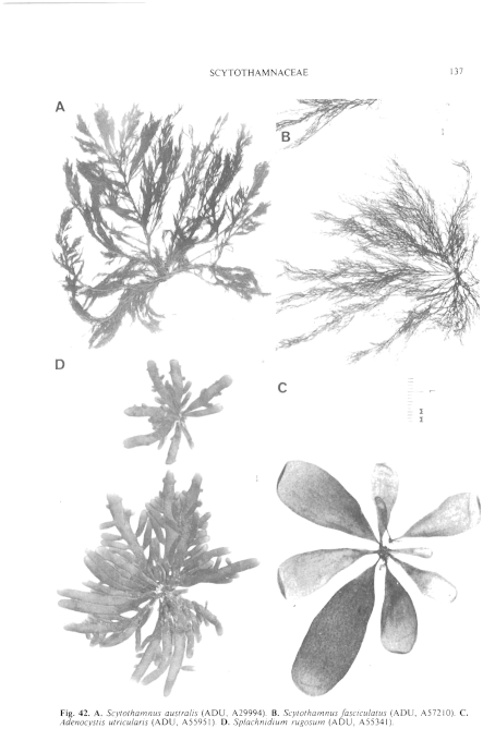
Figure 42 enlarge
Fig. 42. A. Scytothamnus australis (ADU, A29994). B. Scytothamnus fasciculatus (ADU, A57210). C. Adenocystis utricularis (ADU, A55951). D. Splachnidium rugosum (ADU, A55341).
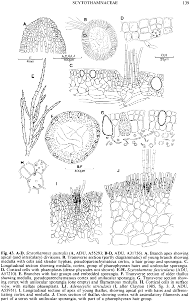
Figure 43 enlarge
Fig. 43. A–D. Scytothamnus australis (A, ADU, A55293; B–D, ADU, A31756). A. Branch apex showing apical (and intercalary) divisions. B. Transverse section (partly diagrammatic) of young branch showing medulla with cells and slender hyphae, pseudoparenchymatous cortex, a hair group and sporangia. C. Longitudinal section showing medulla, cortex, group of phaeophycean hairs and unilocular sporangia. D. Cortical cells with phaeoplasts (dense physodes not shown). E–H. Scytothamnus fasciculatus (ADU, A57210). E. Branches with hair groups and embedded sporangia. F. Transverse section of older thallus showing medulla, pseudoparenchymatous cortex and unilocular sporangia. G. Transverse section showing cortex with unilocular sporangia (one empty) and filamentous medulla. H. Cortical cells in surface view, with stellate phaeoplasts. I,J. Adenocystis utricularis (I, after Clayton 1985, fig. 3; J. ADU, A55951). I. Longitudinal section of apex of young thallus, showing apical pit with hairs and differentiating cortex and medulla. J. Cross section of thallus showing cortex with assimilatory filaments and part of a sorus with unilocular sporangia, with part of a phaeophycean hair group.

|
Email Contact: State Herbarium of South Australia |

|