|
|
|
|
|
|||||||||||
|
Electronic Flora of South Australia Species Fact Sheet
Phylum Phaeophyta – Order Chordariales – Family Myrionemataceae
Thallus medium brown, regularly discoid and domed with the margins adherent to the host and the disc centre becoming free, 0.2–1.0 mm across, epiphytic on Zostera. Basal layer (Fig. 16D–F) monostromatic, of subdichotomous, closely adjacent, filaments radiating from a central group of 4 cells; cells (8–) 12–20 µm long, 4–8 µm broad and 4–6 µm high. Erect assimilatory filaments (Fig. 16F) relatively few, scattered, 30–65 µm and 3–5 cells high, with the cells 10–12 µm long and (3–) 4–7 µm in diameter throughout; phaeoplasts probably 1–3 per cell. Ascocysts (Fig. 16F) frequent, arising from cells of the basal layer, cylindrical to slightly clavate and 2–3 times as high as erect assimilatory filaments, thick walled with hyaline contents, 60–120 µm high and 8–12 (–15) µm in diameter. Hairs (Fig. 16E) phaeophycean, numerous, scattered, 14–20 µm in diameter, with a narrower basal cell and a basal collar.
Reproduction: Plurilocular sporangia (Fig. 16E) filiform, sessile on cells of the basal layer, uniseriate to biseriate with 16–20 locules, opening by a terminal pore, (30–) 45–70 (–120) long and (5–) 7–12 ,,rm in diameter. Unilocular sporangia (Fig. 16F) on separate plants, ovoid, sessile on cells of the basal layer, 40–65 µm long and 20–30 µm in diameter.
Type from the Onkaparinga Estuary, S. Aust., on blades of Zostera muelleri (Thomas, 28.vi.1977; grown in culture); holotype in ADU, A48142.
Distribution: Only known from the Onkaparinga Estuary, S. Aust. (type and Skinner, 1.vii.1977; ADU, A48143).
Taxonomic notes: Myrionema latipilosum is named after its very broad hairs, and belongs to the M. balticum-M. magnusii-M. orbiculare group (Loiseaux 1967b, p. 562) which have prominent ascocysts. It differs from these species in having broader hairs, very long ascocysts, and long, filiform plurilocular sporangia. M. latipilosum has been studied mainly in culture, where it developed more conspicuously on the Zostera blades than was apparent in field collections.
Skinner (1981) showed that the life history of M. latipilosum is probably direct from plurilocular sporangia, but the growth of zooids from unilocular sporangia was not observed.
References:
LOISEAUX, S. (1967b). Recherches sur les cycles de développement des Myrionématacées (Phéophycées). I–II. Hécatonématées et Myrionématées. Rev. Gén. Bot. 74, 529–577, Plates 1, 2.
SKINNER, S. (1981). The taxonomy, morphology and reproduction of the Myrionemaceae, Elachistaceae, Corynophlaeaceae and Giraudyaceae (Phaeophyceae) in southern Australia. Ph. D. thesis, University of Adelaide, unpublished.
The Marine Benthic Flora of Southern Australia Part II complete list of references.
Publication:
Womersley, H.B.S. (14 December, 1987)
The Marine Benthic Flora of Southern Australia
Part II
©Board of the Botanic Gardens and State Herbarium, Government of South Australia
Illustration in Womersley Part II, 1997: FIG. 16 D–F.
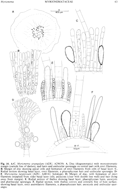
Figure 16 enlarge
Fig. 16. A–C. Myrionema strangulans (ADU, A29639). A. Disc (diagrammatic) with monostromatic margin (outside line of dashes), and hairs and unilocular sporangia on central part with erect filaments. B. Margin of disc showing apical cells and formation of erect filaments from cells of basal layer. C. Radial section showing basal layer, erect filaments, a phaeophycean hair and unilocular sporangia. D–F. Myrionema latipilosum (ADU, A48142, holotype). D. Margin of disc, with formation of erect filaments (stippled) from older basal layer cells, ascocysts (clear with double line wall) and hair (lying away from margin). E. Radial section of thallus showing basal layer, phaeophycean hairs, ascocysts and plurilocular sporangia. F. Radial section, with thallus convex and centrally free of host surface, showing basal layer, erect assimilatory filaments, a phaeophycean hair, ascocysts and unilocular sporangia.

|
Email Contact: State Herbarium of South Australia |

|