|
|
|
|
|
|||||||||||
|
Electronic Flora of South Australia Species Fact Sheet
Phylum Phaeophyta – Order Chordariales – Family Leathesiaceae
Thallus (Fig. 26D) brown, pulvinate to globose, mucoid, 0.5–2.5 mm across, epiphytic on ramuli of Cystophora monilifera, with only slight basal penetration of the host, an extensive medulla and free, widely separated, determinate cortical filaments. Basal filaments forming an entangled layer on and just above the host meristoderm, with short cells adherent to the meristoderm and only slight penetration of filaments between the meristoderm cells. Medulla (Fig. 26D,E) extensive, 300–1000 µm and (8–) 10–25 cells high, dense, with cells ovoid below and 12–15 in diameter, cylindrical above and 7–12 µm in diameter, L/B 3–5, culminating in a dense stratum of plurilocular sporangia. Determinate cortical filaments (Fig. 26D,E) arising from basal filaments and extending unbranched through the medulla, separated, free, cylindrical, 500–1000 (–1400) µm and 20–60 (–80) cells long, with cells thick walled, 7–10 µm in diameter and L/B 1–3. Phaeoplasts (Fig. 26F) several (4–8) per cell, discoid, apparently without a pyrenoid. Phaeophycean hairs frequent, arising from lower medullary cells and extending through the medulla, 8–12 (–14) µm in diameter.
Reproduction: Plurilocular sporangia (Fig. 26E) forming a dense stratum on slender, corymbose branches from upper medullary cells, filiform, simple or once branched, uniseriate or occasionally biseriate with oblique walls, 30–60 (–70) µm long with 8–16 (–24) locules, 5–8 µm in diameter. Occasional short plurilocular sporangia (Fig. 26G) also occur on the mid cells of the determinate cortical filaments, elongate-conical, usually 10–20 µm and 4–8 locules long, 5–6 µm in diameter.
Type from Aldinga, S. Aust., on Cystophora monilifera, drift (Skinner, 14.x.1977); holotype in ADU, A48582.
Distribution: Only known from the type collection.
Taxonomic notes: M. filiformis is a distinctive species of Myriactula with its well-developed medulla, only slight penetration of the host, and long, cylindrical, determinate cortical filaments. It is most closely related to M. caespitosa but differs in its very slight host penetration, the more extensive medulla development, and in the similarity in form to Corynophlaea. From this genus it differs in having separated, largely free, cortical filaments, rather than the dense layer of closely associated filaments, embedded in mucilage, in Corynophlaea.
References: The Marine Benthic Flora of Southern Australia Part II
Publication:
Womersley, H.B.S. (14 December, 1987)
The Marine Benthic Flora of Southern Australia
Part II
©Board of the Botanic Gardens and State Herbarium, Government of South Australia
Illustration in Womersley Part II, 1997: FIG. 26 D–G.
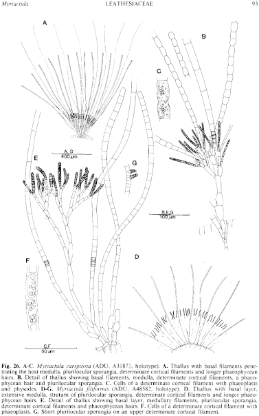
Figure 26 enlarge
Fig. 26. A–C. Myriactula caespitosa (ADU, A3187.±, holotype). A. Thallus with basal filaments penetrating the host medulla, plurilocular sporangia, determinate cortical filaments and longer phaeophycean hairs. B. Detail of thallus showing basal filaments, medulla, determinate cortical filaments, a phaeophycean hair and plurilocular sporangia. C. Cells of a determinate cortical filament with phaeoplasts and physodes. D–G. Myriactula filiformis (ADU, A48582, holotype). D. Thallus with basal layer, extensive medulla, stratum of plurilocular sporangia, determinate cortical filaments and longer phaeophycean hairs. E. Detail of thallus showing basal layer, medullary filaments, plurilocular sporangia, determinate cortical filaments and phaeophycean hairs. F. Cells of a determinate cortical filament with phaeoplasts. G. Short plurilocular sporangia on an upper determinate cortical filament.

|
Email Contact: State Herbarium of South Australia |

|