|
|
|
|
|
|||||||||||
|
Electronic Flora of South Australia Species Fact Sheet
Phylum Phaeophyta – Order Cutleriales – Family Cutleriaceae
Selected citations: Harvey 1847: pl. 75. Kuckuck 1899: 95, pls 7, 8; 1929: 15, figs 5–7. Kylin 1947: 33, pl. 3 fig. 8: Womersley 1967: 207.
Thallus (gametophyte) (Fig. 94A) medium to dark brown, 5–25 (–35) cm long, much branched and often proliferous following damage, with one to several compressed to flat fronds arising from a small rhizoidal holdfast 0.5–2 (–3) mm across and long; epilithic or epiphytic. Growth trichothallic (Fig. 95A) at the base of a tuft of uniseriate apical filaments to each branch; filaments 12–20 mm in diameter, cells L/B (0.3–) 0.5–1 below where meristematic, 2–4 above. Fronds irregularly branched at intervals of mostly 1–3 cm, essentially complanate but branches coming to lie in other planes, 2–8 (–12) mm broad below, 0.5–2 mm broad above, (80–) 100–200 µm broad just below apical tufts, tapering gently or conspicuously from the broader base (or occasionally from the central thallus). Structure 0.7–1.2 mm thick, with a small-celled cortex and large, irregular celled medulla 4–6 (–8) cells thick (Fig. 95C), the 2–4 inner cells being markedly larger; cortical cells angular in surface view, more or less in rows, 6–10 (–12) µm across. Phaeophycean hairs (Fig. 95B) scattered, single or in small clusters, 8–12 µm in diameter.
Sporophyte thallus (Aglaozonia stage) dark brown, dorsiventral, prostrate, flabellate and lobed, with a margin of apical cells (Fig. 95E) segmenting transversely and in the surface plane, becoming 8–10 cells and 120–180 µm thick in mature parts (Fig. 95F), with concentric lines or tufts of phaeophycean hairs on the upper, small-celled cortex and uniseriate rhizoids (Fig. 95F) with digitate holdfast cells from the slightly larger celled lower cortex.
Reproduction: Gametophyte dioecious, bearing surface clusters (Fig. 95B,C) of pedicellate plurilocular gametangia on branched filaments, with some branches extending as phaeophycean hairs; female gametangia (Fig. 95D) ovoid, (40–) 50–80 µm long and 25–30 µm in diameter, with (8–) 16–32 relatively large locules (mostly 4 x 2 locules in side view) and relatively large gametes; male gametangia (unknown in Australia) elongate, with numerous locules and relatively small gametes. Sporophyte with surface sori of unilocular sporangia (Fig. 95F), 40–50 µm long and 10–16 µm in diameter, producing 8 (–32) motile zoospores.
Type from Yarmouth, England (Turner); not located in K or BM.
Selected specimens: (gametophytes): Cockburn Sound, W. Aust., drift (Allender, 25.viii.1966; ADU, A30702). Frenchman Bay, Albany, W. Aust., drift (Parsons, 18.xi.1968; ADU, A33231). Port Turton, S. Aust., 3–5 m deep on piles and rock (Kraft, 17.ix.1973; ADU, A44019). American R. inlet, Kangaroo I., S. Aust., 2–4 m deep on Halophila (Kraft, 2.xii.1971; ADU,
Distribution: Widely distributed in temperate waters.
In Australia, known from Cockburn Sound, W. Aust. to Burraneer, Sydney, N.S.W. and around Tasmania; collected in all months but most frequently in spring and early summer.
Taxonomic notes: A41140) and drift (Womersley, 31.x.1966; ADU, A30832-"Marine Algae of southern Australia" No. 35). Port Elliot, S. Aust., drift (Pocock & Womersley, 20.i.1960; ADU, A23998). Robe, S. Aust., drift in Lake Butler (Womersley, 16.x.1985; ADU, A57020-"Marine Algae of southern Australia" No. 35a). Portland, Vic., drift ( Womersley, 3.ix.1981; ADU, A55373). Geelong, Vic. (Harvey, Alg. Aust. Exsicc. 76; MEL, 15796–7). Swan Bay, Port Philip, Vic., 1 m deep (Watson, 6.x.1973; ADU, A44142). Crawfish Rock, Westernport Bay, Vic., 5–10 m deep ( Watson, 29.viii.1971; ADU, A39379). Triabunna, Tas., sublittoral (Cribb 147.6, 11.vi.1951; ADU, A16358). Burraneer, Sydney, N.S.W., dredged (Levring, 11.xi.1947; ADU, A56002).
Sporophyte (Aglaozonia stage). Williamstown, Vic., 1–2 m deep (Kraft 6599 & Ricker, 26.vii.1978; ADU, A54229).
Cutleria multifida is largely confined to sheltered bays or inlets where it can be locally common. The only record of the Aglaozonia stage is that of Kraft, and Allender & Kraft (1983, p. 122) report it as common in mussel beds in the Melbourne region (i.e. near Port Melbourne) and fertile in mid winter.
References:
ALLENDER, B.M. & KRAFT, G.T. (1983). The marine algae of Lord Howe Island (New South Wales): The Dictyotales and Cutleriales (Phaeophyta). Brunonia 6, 73–130.
GREVILLE, R.K. (1830). Algae Britannicae. (Edinburgh.)
HARVEY, W.H. (1847). Phycologia Britannica. Plates 73–144. (Reeve: London.)
KUCKUCK, P. (1899). Beiträge zur Kenntnis der Meeresalgen. 9. Ober den Generationswechsel von Cutleria multifida (Engl. Bot.) Grev. Wiss. Meeresunters. Abt. Helgol., N.F. 3, 95–117.
KUCKUCK, P. (1929). Fragmente einer Monographie des Pheosporeen. Biol. Anst. Helgol. 17, 1–93.
KYLIN, H. (1947). Die Phaeophyceen der Schwedischen Westkiiste. Acta Univ. lund. N.F. Avd. 2, 43(4), 1–99, Plates 1–18.
The Marine Benthic Flora of Southern Australia Part II complete list of references.
Publication:
Womersley, H.B.S. (14 December, 1987)
The Marine Benthic Flora of Southern Australia
Part II
©Board of the Botanic Gardens and State Herbarium, Government of South Australia
Illustrations in Womersley Part II, 1997: FIGS 94A, 95.
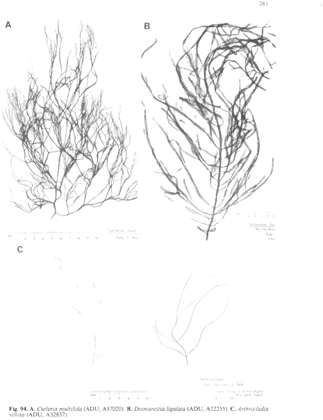
Figure 94 enlarge
Fig. 94. A. Cutleria multifida (ADU, A57020). B. Desmarestia ligulata (ADU, A22255). C. Arthrocladia villosa (ADU, A52837).
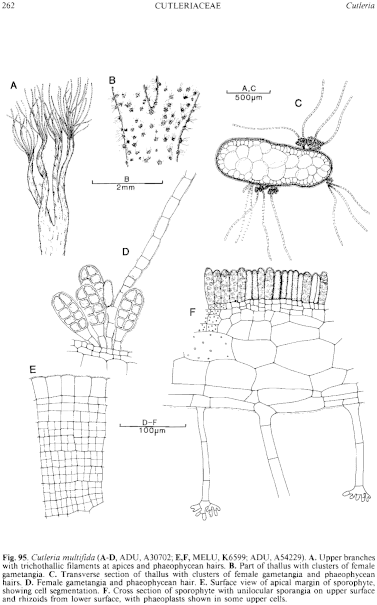
Figure 95 enlarge
Fig. 95. Cutleria multifida (A–C, ADU, A30702; E,F, MELU, K6599; ADU, A54229). A. Upper branches with trichothallic filaments at apices and phaeophycean hairs. B. Part of thallus with clusters of female gametangia. C. Transverse section of thallus with clusters of female gametangia and phaeophycean hairs. D. Female gametangia and phaeophycean hair. E. Surface view of apical margin of sporophyte, showing cell segmentation. F. Cross section of sporophyte with unilocular sporangia on upper surface and rhizoids from lower surface, with phaeoplasts shown in some upper cells.

|
Email Contact: State Herbarium of South Australia |

|