|
|
|
|
|
|||||||||||
|
Electronic Flora of South Australia Species Fact Sheet
Phylum Phaeophyta – Order Ectocarpales
Selected citations: Clayton 1974: 749, fig. 1. King & Ducker 1971: 115. Kornmann & Sahling 1977: 115, fig. 60. Knoepffler-Péguy 1974: 44, figs 1–7, pl. 1. Tanaka & Chihara 1977: 249, fig. 2.
Synonyms
Ectocarpus crinitus Carmichael ex Harvey. Harvey 1850: pl. 330.
A. pusilla (Griffiths & Harvey) Bornet. Blomquist 1955: 46, figs 1–10. Hamel 1931: 75, fig. 22.
Ectocarpus pusillus Griffiths. Bornet 1891: 356, pl. 7 figs 1–5. Harvey 1848: pl. 153.
Thallus (Fig. 10A) medium brown, irregularly branched, forming loose tangled masses to 8 cm long attached to other algae by recurved branchlets, with long branches (Fig. 11A) bearing shorter laterals from centrally on the parent cell, with rounded end cells or tapering slightly into false hairs; short, straight to curved laterals are frequent on older filaments. Filaments (Fig. 11A,B) 24–34 µm in diameter below with cells L/B (0.5–) 1–2 (–3), decreasing to 15–20 µm in diameter in branchlets with cells L/B 0.5–2 (–4), and then to 6–10 µm in diameter in false hairs with cells L/B 4–6. Growth diffuse, sometimes with meristematic regions at the base of laterals (Fig. 11B). Cells (Fig. 11C,D) with numerous discoid phaeoplasts, each with a pyrenoid, and centrally aggregated small physodes.
Reproduction: Plurilocular organs usually sessile (Fig. 11C) with large locules, tapering, 70–120 µm long and 30–40 µm broad; those with small locules (antheridia ?) on different filaments, tapering, usually reflexed forwards (Fig. 11D), 40–80 µm long and 16–28 µm in diameter. Unilocular sporangia (Fig. 11E) terminal on a short branch or single pedicel cell, subspherical, 40–60 µm in diameter. Monosporangia (Fig. 11C) sessile, 20–28 µm in diameter. All reproductive organs usually centrally situated on parent cell.
Type from Appin, Scotland, in BM.
Selected specimens: Granite I., Victor Harbor, S. Aust., upper sublittoral (Clayton, 23.ii.1974; MUCV, 502, ADU, A44771). "The Granites", 16 km N. of Kingston, S. Aust., lower eulittoral, shaded (Womersley, 24.ii.1984; ADU, A54824). Nora Creina, S. Aust., upper sublittoral (Clayton, 17.ii.1974; MUCV, 501, ADU, A44772). Point Bunbury, Apollo Bay, Vic., lower eulittoral pool (Womersley, 12.iv.1959; ADU, A22658). Sorrento, Vic., dredged 10 m deep (Womersley, 7.iv.1969; ADU, A22770). Nambucca Heads, N.S.W. (Clayton, 26.v.1971; MELU, 21029).
Distribution: Europe, western North Atlantic, Japan.
In southern Australia, from Granite I., Victor Harbor, S. Aust. to Nambucca Heads, N.S.W.
Taxonomic notes: Kornmann (1953) considers that Acinetospora is the diploid phase and Feldmannia lebelii (or F. padinae) is the haploid phase of a single species. Both phases occur on southern Australian coasts and they may well be connected but the nature of the various reproductive organs awaits detailed study.
References:
BLOMQUIST, H.L. (1955). Acinetospora Born. new to North America. J. Elisha Mitchell Sci. Soc. 71, 46–49.
BORNET, E. (1891). Note sur quelques Ectocarpus. Bull. Soc. Bot. Fr. 38, 353–372.
CLAYTON, M.N. (1974). Studies on the development, life history and taxonomy of the Ectocarpales (Phaeophyta) in southern Australia. Aust. J. Bot. 22, 743–813.
HAMEL, G. (1931). Phéophycées de France. Fasc. I, pp. 1–80. (Paris.)
HARVEY, W.H. (1848). Phycologia Britannica. Plates 145–216. (Reeve: London.)
HARVEY, W.H. (1850). Phycologia Britannica. Plates 253–354. (Reeve: London.)
KING, R.J. & DUCKER, S. (1971). In King, R.J., Black, J.H. & Ducker, S. Intertidal ecology of Port Phillip Bay, with systematic lists of plants and animals. 3. Flora of the intertidal region, pp. 112–128. Mem. Nat. Mus. Vic. 32, 93–128.
KNOEPFFLER-PÉGUY, M. (1974). Le genre Acinetospora Bornet 1891 (Phaeophyceae-Ectocarpales). Vie et Milieu, Sér. A, 24, 43–72.
KORNMANN, P. & SAHLING, P.-H. (1977). Meeresalgen von Helgoland. Benthische Braun und Rotalgen. Helgol. wiss. Meeresunters. 29, 1–292.
KORNMANN, P. (1953). Der Formenkreis von Acinetospora crinita (Carm.) nov. comb. Helgol. wiss. Meeresunters. 4, 205–224.
TANAKA, J. & CHIHARA, M. (1977). Notes on algae in Japan and adjacent waters (1). J. Jap. Bot. 52, 245–253.
The Marine Benthic Flora of Southern Australia Part II complete list of references.
Publication:
Womersley, H.B.S. (14 December, 1987)
The Marine Benthic Flora of Southern Australia
Part II
©Board of the Botanic Gardens and State Herbarium, Government of South Australia
Illustrations in Womersley Part II, 1997: FIGS 10A, 11 A–E.
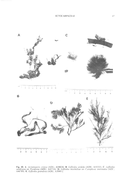
Figure 10 enlarge
Fig. 10. A. Acinetospora crinita (ADU, A54824). B. Giffordia sordida (ADU, A55332). C. Giffordia sandriana on Posidonia (ADU, A42716). D. Giffordia mitchelliae on Cystophora intermedia (ADU, A46789). E. Giffordia granulosa (ADU, A39491).
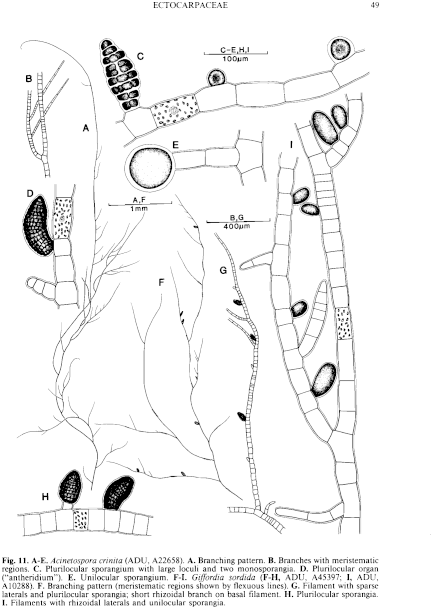
Figure 11 enlarge
Fig. 11. A–E. Acinetospora crinita (ADU, A22658). A. Branching pattern. B. Branches with meristematic regions. C. Plurilocular sporangium with large loculi and two monosporangia. D. Plurilocular organ ("antheridium"). E. Unilocular sporangium. F–I. Giffordia sordida (F–H, ADU, A45397; I, ADU, A10288). F. Branching pattern (meristematic regions shown by flexuous lines). G. Filament with sparse laterals and pluriLocular sporangia; short rhizoidal branch on basal filament. H. Plurilocular sporangia. I. Filaments with rhizoidal laterals and unilocular sporangia.

|
Email Contact: State Herbarium of South Australia |

|